


Effect of transient scrotal hyperthermia on sperm parameters, seminal plasma biochemical markers, and oxidative stress in men
- PMID: 25652627
- PMCID: PMC4492061
- DOI: 10.4103/1008-682X.146967
Effect of transient scrotal hyperthermia on sperm parameters, seminal plasma biochemical markers, and oxidative stress in men
Abstract
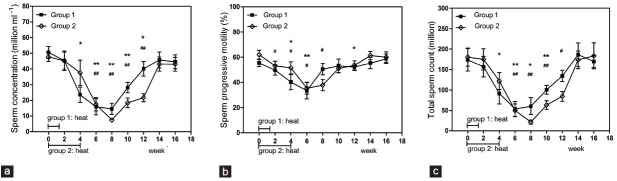
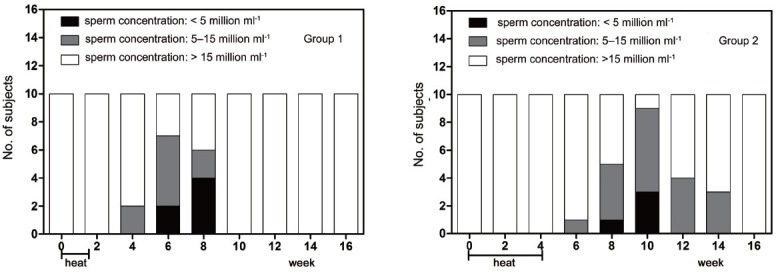
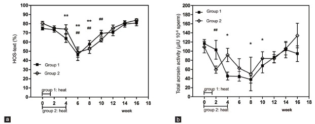
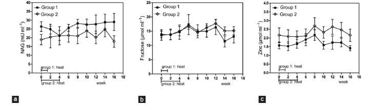
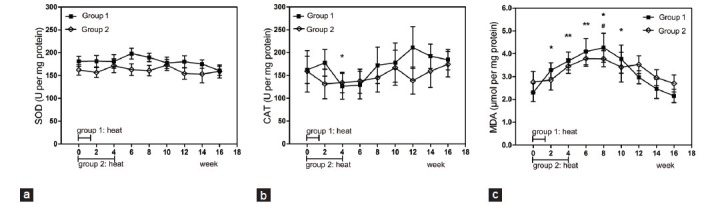
In this experimental prospective study, we aimed to analyze the effect of transient scrotal hyperthermia on the male reproductive organs, from the perspective of sperm parameters, semen plasma biochemical markers, and oxidative stress, to evaluate whether different frequencies of heat exposure cause different degrees of damage to spermatogenesis. Two groups of volunteers (10 per group) received testicular warming in a 43°C water bath 10 times, for 30 min each time: group 1: 10 consecutive days; group 2: once every 3 days. Sperm parameters, epididymis and accessory sex gland function, semen plasma oxidative stress and serum sex hormones were tested before treatment and in the 16-week recovery period after treatment. At last, we found an obvious reversible decrease in sperm concentration (P = 0.005 for Group 1 and P= 0.008 for Group 2 when the minimums were compared with baseline levels, the same below), motility (P = 0.009 and 0.021, respectively), the hypoosmotic swelling test score (P = 0.007 and 0.008, respectively), total acrosin activity (P = 0.018 and 0.009, respectively), and an increase in the seminal plasma malondialdehyde concentration (P = 0.005 and 0.017, respectively). The decrease of sperm concentration was greater for Group 2 than for Group 1 (P = 0.031). We concluded that transient scrotal hyperthermia seriously, but reversibly, negatively affected the spermatogenesis, oxidative stress may be involved in this process. In addition, intermittent heat exposure more seriously suppresses the spermatogenesis compared to consecutive heat exposure. This may be indicative for clinical infertility etiology analysis and the design of contraceptive methods based on heat stress.
Figures
Similar articles
- Improvement of semen quality by nocturnal scrotal cooling and moderate behavioural change to reduce genital heat stress in men with oligoasthenoteratozoospermia.Reproduction. 2001 Apr;121(4):595-603. doi: 10.1530/rep.0.1210595.Reproduction. 2001.PMID: 11277880
- Changes in miRNA levels of sperm and small extracellular vesicles of seminal plasma are associated with transient scrotal heat stress in bulls.Theriogenology. 2021 Feb;161:26-40. doi: 10.1016/j.theriogenology.2020.11.015. Epub 2020 Nov 27.Theriogenology. 2021.PMID: 33278692
- Analysis of human sperm DNA fragmentation index (DFI) related factors: a report of 1010 subfertile men in China.Reprod Biol Endocrinol. 2018 Mar 14;16(1):23. doi: 10.1186/s12958-018-0345-y.Reprod Biol Endocrinol. 2018.PMID: 29540184Free PMC article.
- Review: Testicular vascular cone development and its association with scrotal thermoregulation, semen quality and sperm production in bulls.Animal. 2018 Jun;12(s1):s133-s141. doi: 10.1017/S1751731118001167.Animal. 2018.PMID: 29882506Review.
- Influence of genital heat stress on semen quality in humans.Andrologia. 2007 Dec;39(6):203-15. doi: 10.1111/j.1439-0272.2007.00794.x.Andrologia. 2007.PMID: 18076419Review.
Cited by
- Association between terbinafine hydrochloride and sperm DNA fragmentation - case report.JBRA Assist Reprod. 2022 Apr 17;26(2):366-370. doi: 10.5935/1518-0557.20210093.JBRA Assist Reprod. 2022.PMID: 34751019Free PMC article.
- The Importance of Oxidative Stress in Determining the Functionality of Mammalian Spermatozoa: A Two-Edged Sword.Antioxidants (Basel). 2020 Jan 27;9(2):111. doi: 10.3390/antiox9020111.Antioxidants (Basel). 2020.PMID: 32012712Free PMC article.Review.
- Effects of meteorology and lunar cycle on the post-thawing quality of avian sperm.Front Vet Sci. 2024 May 16;11:1394004. doi: 10.3389/fvets.2024.1394004. eCollection 2024.Front Vet Sci. 2024.PMID: 38818498Free PMC article.
- Timescale and colony-dependent relationships between environmental conditions and plasma oxidative markers in a long-lived bat species.Conserv Physiol. 2020 Sep 14;8(1):coaa083. doi: 10.1093/conphys/coaa083. eCollection 2020.Conserv Physiol. 2020.PMID: 33173584Free PMC article.
- Hormonal, chemical and thermal inhibition of spermatogenesis: contribution of French teams to international data with the aim of developing male contraception in France.Basic Clin Androl. 2017 Jan 13;27:3. doi: 10.1186/s12610-016-0047-2. eCollection 2017.Basic Clin Androl. 2017.PMID: 28101363Free PMC article.
References
-
- Thonneau P, Ducot B, Bujan L, Mieusset R, Spira A. Effect of male occupational heat exposure on time to pregnancy. Int J Androl. 1997;20:274–8. - PubMed
-
- Hjollund NH, Bonde JP, Jensen TK, Olsen J. Diurnal scrotal skin temperature and semen quality. The Danish First Pregnancy Planner Study Team. Int J Androl. 2000;23:309–18. - PubMed
-
- Garolla A, Torino M, Sartini B, Cosci I, Patassini C, et al. Seminal and molecular evidence that sauna exposure affects human spermatogenesis. Hum Reprod. 2013;28:877–85. - PubMed
Publication types
MeSH terms
Substances
LinkOut - more resources
Full Text Sources
Medical
